
Radiology’s Role in Neuromorphology: Betbhai9 whatsapp number, Play exch.in, Lotus365.win new id
betbhai9 whatsapp number, play exch.in, lotus365.win new id: Radiology’s Role in Neuromorphology
Are you curious about how radiology plays a crucial role in the field of neuromorphology? As technology continues to advance, radiology has become an invaluable tool in studying the structure and function of the brain. In this blog post, we will explore the significance of radiology in neuromorphology and how it is shaping our understanding of the brain.
The Basics of Neuromorphology
Neuromorphology is the study of the shape and structure of the nervous system, particularly the brain. It involves analyzing the morphology of neurons, glial cells, and other components of the nervous system to better understand their function. By studying the intricate details of the brain’s anatomy, researchers can gain insights into how it processes information, controls behavior, and responds to various stimuli.
Radiology’s Contribution to Neuromorphology
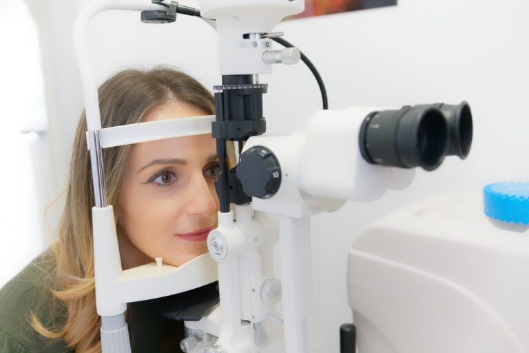
Radiology, specifically techniques like magnetic resonance imaging (MRI) and computed tomography (CT), plays a vital role in neuromorphology. These imaging modalities allow researchers and clinicians to visualize the brain in great detail, capturing images of its structure and detecting abnormalities that may be present. MRI, in particular, is an excellent tool for studying the brain’s anatomy as it provides high-resolution images without the use of harmful radiation.
By utilizing radiology techniques, researchers can map out the brain’s regions, identify connections between different areas, and track changes over time. This information is crucial for understanding how the brain develops, functions, and responds to injury or disease. Radiology also plays a significant role in the diagnosis and treatment of neurological disorders, providing valuable insights that can guide patient care.
The Impact of Advanced Imaging Techniques
Recent advancements in imaging technology have revolutionized the field of neuromorphology. Techniques such as diffusion tensor imaging (DTI) and functional MRI (fMRI) allow researchers to study the brain’s white matter tracts and neural activity in real-time. These imaging modalities provide a deeper understanding of brain function and connectivity, shedding light on complex neurological processes.
DTI, for example, enables researchers to track the pathways of white matter fibers in the brain, revealing how different regions are connected and how information is transmitted between them. This information is invaluable for studying conditions like traumatic brain injury, stroke, and neurodegenerative diseases, where disruptions in neural connectivity play a significant role.
Similarly, fMRI allows researchers to observe changes in blood flow and oxygen levels in the brain as a person performs various tasks or experiences stimuli. By studying these patterns of brain activity, researchers can gain insights into cognitive processes, emotion regulation, and sensory perception. fMRI has been instrumental in studying conditions like schizophrenia, depression, and Alzheimer’s disease, providing valuable information for diagnosis and treatment.
Common Applications of Radiology in Neuromorphology
Radiology is used in various research and clinical settings to study the brain’s structure and function. Some common applications of radiology in neuromorphology include:
– Diagnosing neurological disorders: Radiology is often used to detect abnormalities in the brain, such as tumors, lesions, and hemorrhages, that may indicate the presence of a neurological disorder. Imaging techniques can help clinicians make accurate diagnoses and develop treatment plans for patients.
– Monitoring brain development: Radiology is used to track changes in the brain’s structure over time, allowing researchers to study how the brain develops from infancy to adulthood. This information is crucial for understanding normal brain growth and identifying deviations that may indicate developmental disorders.
– Studying cognitive processes: Radiology is used in cognitive neuroscience research to study how the brain processes information, makes decisions, and controls behavior. Imaging modalities like fMRI allow researchers to observe patterns of brain activity associated with specific cognitive tasks, providing insights into how the brain functions.
– Investigating brain connectivity: Radiology is used to study the connectivity of the brain’s neural networks, mapping out the pathways that connect different regions. Understanding these connections is essential for studying how information is processed, integrated, and relayed within the brain.
– Assessing treatment outcomes: Radiology is used to evaluate the effectiveness of treatments for neurological disorders, such as surgeries, medications, and behavioral therapies. Imaging techniques can track changes in the brain before and after treatment, helping clinicians assess the impact of interventions on brain structure and function.
FAQs
1. What is the difference between MRI and CT scans in studying the brain?
MRI uses magnetic fields and radio waves to create detailed images of the brain’s structure, while CT scans use X-rays to generate cross-sectional images. MRI is preferred for studying soft tissues, such as the brain, as it provides higher resolution images without exposing the patient to ionizing radiation.
2. How is DTI used to study white matter tracts in the brain?
DTI measures the diffusion of water molecules in the brain’s white matter tracts, allowing researchers to track the pathways of neural fibers. By studying these pathways, researchers can map out the connectivity of different brain regions and study how information is transmitted between them.
3. What are some common neurological disorders that benefit from radiology imaging?
Conditions like stroke, traumatic brain injury, Alzheimer’s disease, multiple sclerosis, and brain tumors are commonly studied using radiology imaging techniques. These modalities help clinicians diagnose these disorders, track disease progression, and assess treatment outcomes.
4. How has fMRI advanced our understanding of brain function?
fMRI has allowed researchers to observe changes in brain activity in real-time as a person performs tasks or experiences stimuli. By studying these patterns of brain activity, researchers can gain insights into cognitive processes, emotional responses, and sensory perception, advancing our understanding of how the brain functions.
In conclusion, radiology plays a vital role in the field of neuromorphology by providing detailed images of the brain’s structure and function. Through advanced imaging techniques like MRI, DTI, and fMRI, researchers can study the brain’s anatomy, connectivity, and cognitive processes, leading to new insights into neurological disorders and brain function. As technology continues to evolve, radiology will continue to shape our understanding of the brain and contribute to advancements in neuroscience.